More Stem Cells—or Better Stem Cells? How Laboratory Expansion Can Reduce Cellular Potency *Educational Purposes Only*
In regenerative medicine, patients are frequently impressed by large cell counts. Some international clinics, including programs operating in Mexico, advertise infusions containing hundreds of millions—or even more than a billion—culture-expanded mesenchymal stromal cells.
At first glance, more cells sounds better. The biology, however, is more complicated.
Mesenchymal stromal cells, commonly called mesenchymal stem cells or MSCs, cannot be multiplied indefinitely without changing them. Producing very large doses requires laboratories to repeatedly grow, divide, harvest, and replate the cells. Each cycle is called a passage.
Research shows that serial passage can progressively alter MSC proliferation, differentiation potential, gene expression, mitochondrial function, secretory behavior, immune-modulating activity, and genomic stability.¹–⁶ This does not mean every culture-expanded MSC product is ineffective. It does mean that cell count alone is an inadequate measure of cell quality.
What Is Cell Expansion?
MSCs occur in relatively small numbers within bone marrow, adipose tissue, umbilical cord, placenta, and other tissues. To create larger doses, laboratories place isolated cells into culture media that encourage proliferation.
Once the cells cover enough of the culture surface, they are detached and transferred into new vessels. These transfers are designated P1, P2, P3, and so forth.
Each passage increases the number of cells, but it also exposes them to continued replication, artificial culture surfaces, oxidative stress, enzymatic detachment, and loss of the signals normally provided by their native tissue environment.
The real manufacturing challenge is not simply producing more cells. It is producing enough cells while preserving their biologic identity and functional potency.
Passage Number Is Only Part of the Story
Passage number is useful but incomplete. Two laboratories may label cells as P3 even though the cells have undergone different numbers of actual divisions.
More informative manufacturing measurements include:
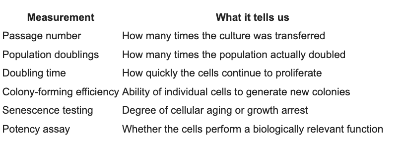
A product described only as containing “500 million stem cells” provides almost none of this information.
Replicative Senescence
MSCs are not immortal. With repeated division, they gradually develop replicative senescence.
Senescent cells may remain alive, but they no longer behave like young, highly functional progenitor cells. They become larger and flatter, divide more slowly, accumulate oxidative and mitochondrial damage, and change the signals they release.⁴
Common findings include:
increased senescence-associated β-galactosidase;
increased p16, p21, and p53 activity;
reduced proliferative capacity;
telomere shortening;
mitochondrial dysfunction;
increased reactive oxygen species;
reduced colony formation;
altered differentiation potential;
changes in cytokine and growth-factor secretion.
The deterioration is not necessarily limited to extremely late passages. Molecular evidence of senescence can appear in relatively early cultures, depending on donor age, tissue source, cell density, oxygen concentration, media, and manufacturing conditions.¹,⁴,⁵
Does Every Passage Make the Cells Less Effective?
Serial expansion generally creates a progressive risk of functional decline, but there is no scientifically valid formula stating that each passage reduces potency by a fixed percentage.
The overall relationship is better represented qualitatively:
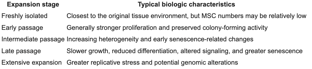
Passage number should therefore not be converted into an assumed potency percentage unless the manufacturer has validated that relationship using a clinically relevant assay.
Early-Passage Cells Can Already Be Different
Jiang and colleagues compared freshly isolated bone-marrow cells with passage-3 MSCs in a cartilage-defect model. Although the expanded cells retained conventional MSC characteristics, expansion altered their stemness-related properties and reduced their cartilage-regeneration performance.¹
This illustrates an important limitation of standard cell identification. Expanded MSCs may continue to express markers such as CD73, CD90, and CD105 while having diminished colony formation, trophic signaling, differentiation, homing, or immunomodulatory activity.
Cell identity is not the same as cell potency.
Hoch and Leach similarly described substantial degeneration of progenitor potency during conventional two-dimensional culture expansion.² Yang and colleagues reported passage-associated changes in MSC morphology, gene expression, phenotype, and differentiation capacity.³
The Secretome May Matter More Than Engraftment
The early stem-cell narrative suggested that administered MSCs engraft and directly replace damaged cartilage, tendon, muscle, or bone. Current MSC biology suggests that much of their potential activity may instead come from paracrine signaling.
MSCs release a secretome containing cytokines, chemokines, growth factors, extracellular matrix proteins, extracellular vesicles, and regulatory microRNAs. These signals may influence macrophages, lymphocytes, blood vessels, fibroblasts, and the patient’s resident repair cells.
Senescence changes this secretome. Extensively expanded cells may release fewer desirable trophic signals and more inflammatory factors associated with the senescence-associated secretory phenotype, or SASP.⁴
Consequently, two products containing the same number of viable cells may not have the same biologic activity. Basic viability testing confirms that a cell is alive; it does not prove that the cell remains therapeutically potent.
Genomic Stability Also Matters
Repeated cell division creates opportunities for replication-related genetic changes.
Kim and colleagues used whole-genome sequencing to evaluate MSCs from P1 through P9. They found passage-dependent accumulation of somatic variants, accompanied by longer doubling times, reduced colony formation, telomere shortening, and increasingly senescent morphology.⁶
This does not establish that expanded MSC products inevitably become malignant. It does show why prolonged expansion requires rigorous testing for genomic stability, growth behavior, sterility, identity, and biologic potency.
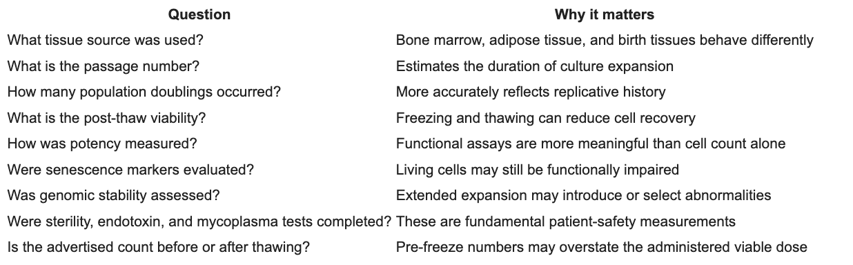
Questions to Ask About High Cell Counts
When a clinic advertises hundreds of millions or billions of MSCs, those cells almost certainly required substantial laboratory expansion. That does not automatically make the product poor, but it should prompt important questions:
Why We Use U.S.-Based Tissue Products
At RegeneZone™, we do not select a biologic product simply because it advertises the largest number of cells. We prioritize traceability, donor screening, tissue processing, manufacturing controls, independent lot testing, and compliance with the applicable U.S. regulatory framework.
We use products supplied by U.S.-based companies working with FDA-registered tissue establishments and operating under the regulations applicable to human cells, tissues, and cellular and tissue-based products, or HCT/Ps. FDA registration and product listing do not mean that a product has been individually approved by the FDA, but they place the establishment within an established federal system governing donor eligibility, communicable-disease controls, recordkeeping, processing, and current good tissue practices. (U.S. Food and Drug Administration)
One of the birth-tissue products we use when clinically appropriate is HylaPure®, supplied by BioXstem. HylaPure is described by the company as a minimally manipulated, DMSO-free, cryopreserved human umbilical-cord tissue allograft. The donated tissue is obtained in the United States from screened, consenting mothers following full-term Caesarean delivery. (BioXstem)
HylaPure should not be confused with a laboratory-expanded MSC drug containing hundreds of millions of culture-proliferated cells. It is a structural human birth-tissue allograft intended to preserve naturally occurring tissue components within the donated umbilical-cord matrix.
This distinction is important. Our objective is not to compete with international clinics by advertising the largest theoretical cell count. Our objective is to select well-characterized, responsibly sourced biologic products and integrate them into a carefully designed regenerative treatment plan.
The Bottom Line
The scientific evidence supports several conclusions:
MSCs change during laboratory expansion.
Repeated passage can increase cellular senescence and functional drift.
Expansion may reduce proliferation, colony formation, differentiation, immunomodulation, and regenerative activity.
Standard MSC surface markers do not establish therapeutic potency.
Later passages may accumulate greater replicative and genomic abnormalities.
The degree of deterioration varies according to donor, tissue source, and manufacturing method.
A very high cell count does not prove that a product is more biologically effective.
The most important question is not simply:
“How many stem cells am I receiving?”
The better questions are:
“Where did the product come from, how was it processed, how extensively were its cells expanded, and what objective testing supports its quality?”
In regenerative medicine, more is not necessarily better. Cellular quality, tissue integrity, manufacturing transparency, and clinical judgment may matter far more than the largest number printed on a treatment brochure.
References
Jiang T, Xu G, Wang Q, et al. In vitro expansion impaired the stemness of early passage mesenchymal stem cells for treatment of cartilage defects. Cell Death & Disease. 2017;8:e2851. doi:10.1038/cddis.2017.215.
Hoch AI, Leach JK. Concise review: Optimizing expansion of bone marrow mesenchymal stem/stromal cells for clinical applications. Stem Cells Translational Medicine. 2014;3(5):643–652. doi:10.5966/sctm.2013-0196.
Yang YHK, Ogando CR, See CW, Chang TY, Barabino GA. Changes in phenotype and differentiation potential of human mesenchymal stem cells aging in vitro. Stem Cell Research & Therapy. 2018;9:131. doi:10.1186/s13287-018-0876-3.
Liu J, Ding Y, Liu Z, Liang X. Senescence in mesenchymal stem cells: Functional alterations, molecular mechanisms, and rejuvenation strategies. Frontiers in Cell and Developmental Biology. 2020;8:258. doi:10.3389/fcell.2020.00258.
Alves-Paiva RM, do Amaral R, Passos GA, et al. Senescence state in mesenchymal stem cells at low passages: Implications in clinical use. Frontiers in Cell and Developmental Biology. 2022;10:858996. doi:10.3389/fcell.2022.858996.
Kim M, Rhee JK, Choi H, et al. Passage-dependent accumulation of somatic mutations in mesenchymal stromal cells during in vitro culture revealed by whole-genome sequencing. Scientific Reports. 2017;7:14508. doi:10.1038/s41598-017-15155-5.